Patient guide + clinician detail

What causes high blood pressure?

High blood pressure usually builds from many forces at once: artery stiffness, salt sensitivity, kidney and hormone signaling, sleep apnea, weight, alcohol, medicines, genetics, age, and cardiovascular disease. The cause matters because the fix is not always just "try harder."

Emergency warning: blood pressure higher than 180/120 mm Hg with chest pain, shortness of breath, back pain, weakness, numbness, vision change, confusion, or trouble speaking can be a hypertensive emergency. Call emergency services. This site is educational and cannot diagnose or treat an individual person.

Choose the language level

Plain language for patients. Medical detail for doctors.

First principle

Hypertension is often silent, but it is not harmless. It is pressure injury happening inside arteries, heart, brain, kidneys, eyes, and blood vessels.

Most people with high blood pressure do not feel it. That is why a careful measurement matters more than symptoms. A single high reading can happen from pain, stress, caffeine, exercise, a full bladder, or poor technique. Hypertension means pressure is persistently high enough to raise risk.

For many adults, high blood pressure is "primary hypertension," meaning there is no single cause. For others, the blood pressure is driven by a specific condition such as kidney disease, sleep apnea, hormone excess, pregnancy, or a medicine.

Hypertension is a continuous cardiovascular risk variable, but classification improves detection, treatment thresholds, and communication. Evaluate technique, out-of-office confirmation, global ASCVD risk, target-organ damage, comorbid CKD/diabetes/HF/CAD/stroke, and secondary causes when suggested by severity, age, resistance, potassium, renal findings, sleep symptoms, or medication history.

Primary hypertension reflects interacting renal sodium handling, RAAS and sympathetic tone, vascular stiffness, endothelial dysfunction, adiposity, insulin resistance, genetics, inflammation, sleep biology, diet, alcohol, and environment. Secondary hypertension should be actively sought when the phenotype is not ordinary.

How high is high?

Blood pressure categories used for most nonpregnant adults.

Category Systolic Diastolic Meaning
Normal Less than 120 and less than 80 Keep measuring over time.
Elevated 120-129 and less than 80 Risk is rising; lifestyle changes matter.
Stage 1 hypertension 130-139 or 80-89 Confirm with repeated readings and risk assessment.
Stage 2 hypertension 140 or higher or 90 or higher Usually needs medication plus lifestyle treatment.
Severe / crisis range Higher than 180 and/or higher than 120 Symptoms or organ injury make this an emergency.

If the top and bottom numbers fall in different categories, the higher category usually determines the level. Individual targets can differ in pregnancy, frailty, kidney disease, diabetes, and established cardiovascular disease.

Basic physiology

Blood pressure is flow multiplied by resistance.

In simple terms, blood pressure depends on how much blood the heart pumps, how tight or stiff the arteries are, how much salt and water the kidneys keep, and which hormones are telling vessels, kidneys, and the heart to speed up or clamp down.

BPBlood pressure
=
Cardiac outputHeart rate x stroke volume
x
Vascular resistanceHow tight and stiff the arterial system is
Aging vessels

Stiff arteries raise systolic pressure

With aging, arteries often lose some stretch. A stiffer aorta and large arteries do not cushion each heartbeat as well, so the top number can climb even when the bottom number is normal or lower.

Arterial stiffening increases pulse wave velocity, wave reflection, pulse pressure, and LV afterload. Isolated systolic hypertension in older adults often reflects large-artery stiffness more than a high-output state.

Resistance

Small arteries set the clamp

Tiny muscular arteries can narrow or relax. When many of them stay narrowed, vascular resistance rises and the heart has to push against a tighter circuit.

Systemic vascular resistance is largely controlled by arteriolar tone, endothelial function, sympathetic activity, angiotensin II, nitric oxide balance, vascular remodeling, and local metabolic signals.

Cardiac output

More flow can raise pressure

Pressure can rise when the heart pumps faster or harder, as with anxiety, pain, stimulants, overactive thyroid, fever, anemia, or adrenaline surges.

CO-driven hypertension may involve increased heart rate, stroke volume, intravascular volume, thyroid excess, catecholamine excess, high-output physiology, or early obesity-related hemodynamics before fixed resistance dominates.

Kidneys

Salt and water control volume

The kidneys decide how much sodium and water to keep. High salt intake, kidney disease, or salt-sensitive biology can expand blood volume and keep pressure high.

Pressure natriuresis, nephron number, tubular sodium handling, CKD, aldosterone signaling, and renal perfusion all affect extracellular volume. Salt sensitivity is more common with older age, CKD, diabetes, metabolic syndrome, and some ancestry groups.

RAAS

Renin, angiotensin, and aldosterone

When kidneys sense low flow, they can release renin. This can lead to angiotensin II, which tightens vessels, and aldosterone, which tells the body to keep salt and water.

The renin-angiotensin-aldosterone system links renal perfusion and sodium balance to vasoconstriction and mineralocorticoid-mediated volume expansion. Renal artery stenosis can drive renin; primary aldosteronism can suppress renin while maintaining hypertension.

Stress hormones

Adrenaline, noradrenaline, and ADH

Epinephrine and norepinephrine can raise heart rate, squeezing strength, and vessel tone. ADH, also called vasopressin, helps the body hold water and can tighten blood vessels in some settings.

Sympathetic activation, catecholamines, vasopressin/ADH, cortisol, thyroid hormone, parathyroid hormone, and sex-steroid or medication effects can shift vascular tone, CO, renal sodium handling, and volume status.

Cause map

The main causes, in plain English.

Etiologic differential, grouped for practical hypertension workup.

01

Primary hypertension: genes, age, arteries, and environment

Most high blood pressure is not caused by one disease. It comes from inherited tendency plus aging arteries, body weight, salt sensitivity, stress biology, low activity, diet, alcohol, and sleep patterns.

Pattern: it often appears gradually and runs in families, but lifestyle and medical treatment can still lower risk.

Primary hypertension is polygenic and multifactorial, with renal sodium retention, arterial stiffness, sympathetic activation, RAAS tone, endothelial dysfunction, adiposity, insulin resistance, inflammation, and social/environmental drivers. Phenotype risk and end-organ effects rather than treating it as benign inevitability.

02

Kidney disease, renal artery stenosis, and salt handling

The kidneys help decide how much salt and water the body keeps. Kidney disease, narrowed kidney arteries, high salt intake, or salt sensitivity can raise blood pressure and make it harder to control.

Clue: abnormal kidney blood tests, protein in urine, swelling, resistant blood pressure, sudden worsening, or a rise in creatinine after some BP medicines may point toward kidney involvement.

Evaluate CKD, albuminuria, renovascular disease including renal artery stenosis/fibromuscular dysplasia, reflux/scarring, glomerular disease, polycystic kidney disease, high sodium intake, and nephrotoxic exposure. Check creatinine/eGFR, electrolytes, urinalysis, UACR, and renal imaging or vascular evaluation when indicated.

03

Sleep apnea and nighttime pressure

Obstructive sleep apnea can repeatedly lower oxygen and trigger stress hormones during sleep. Blood pressure may stay high overnight and be difficult to control during the day.

Clue: loud snoring, witnessed pauses, morning headaches, daytime sleepiness, large neck size, or resistant hypertension.

OSA is common in resistant hypertension and blunted/nocturnal nondipping phenotypes. Screen by symptoms and risk profile; sleep testing and CPAP/weight strategies can support BP control and cardiometabolic risk reduction.

04

Hormone causes: aldosterone, thyroid, cortisol, and adrenaline

Some hormone problems raise blood pressure by holding onto salt, narrowing blood vessels, or speeding up the body. Examples include excess aldosterone, hyperthyroidism, hypothyroidism in some cases, Cushing syndrome, parathyroid disease, and pheochromocytoma, a rare adrenaline-producing tumor.

Clue: low potassium, spells of sweating and palpitations, tremor, heat intolerance, weight loss, very early onset, very severe readings, or blood pressure that needs many medicines.

Consider primary aldosteronism, pheochromocytoma/paraganglioma, Cushing syndrome, hyperthyroidism, hypothyroidism, hyperparathyroidism, acromegaly, congenital adrenal hyperplasia, and other mineralocorticoid excess states. Primary aldosteronism is underdiagnosed and may occur without hypokalemia.

05

Medicines, substances, and supplements

Common products can raise blood pressure: NSAID pain relievers, decongestants, stimulants, some antidepressants, steroids, birth control hormones, licorice, nicotine, cocaine, amphetamines, and heavy alcohol use.

Tip: bring all prescriptions, over-the-counter medicines, supplements, and recreational substances to a blood pressure visit.

Review NSAIDs, sympathomimetics, SNRIs, stimulants, glucocorticoids, calcineurin inhibitors, VEGF inhibitors, erythropoietin, oral contraceptives, testosterone/anabolic steroids, nicotine, alcohol, cocaine/amphetamines, herbal products, and licorice.

06

Weight, insulin resistance, diabetes, and fatty liver biology

Extra body fat, especially around the waist, can increase blood volume, inflammation, insulin resistance, sleep apnea, and kidney salt retention. Diabetes and high blood pressure often travel together.

Important: even modest weight loss, better fitness, and improved sleep can lower pressure for many people.

Adiposity-related hypertension involves sympathetic tone, RAAS activation, sodium retention, OSA, insulin resistance, inflammation, and vascular dysfunction. Treat with BP control plus cardiometabolic risk reduction, not BP numbers alone.

07

Pregnancy-related high blood pressure

High blood pressure during pregnancy or after delivery can be dangerous for the parent and baby. Preeclampsia can involve headaches, vision changes, belly pain, swelling, abnormal labs, or seizures.

Do not wait: pregnancy and postpartum blood pressure problems need urgent medical guidance.

Distinguish chronic hypertension, gestational hypertension, preeclampsia/eclampsia, HELLP, superimposed preeclampsia, and postpartum hypertension. This site's adult category table is not a substitute for obstetric thresholds and protocols.

08

Genetic, vascular, neurologic, and rare causes

Family history is common in primary hypertension, and many genes can each add a small amount of risk. Less common causes include narrowed aorta from birth, adrenal tumors, severe pain, brain pressure, certain autoimmune diseases, and rare single-gene salt-handling conditions.

Clue: young age, sudden onset, arm-leg blood pressure differences, unusual lab results, or severe treatment resistance deserves a deeper look.

Consider coarctation, large-vessel vasculitis, autonomic dysfunction, baroreflex failure, intracranial pathology, polygenic risk, monogenic hypertension, Liddle syndrome, apparent mineralocorticoid excess, medication withdrawal, and hypertensive emergency mimics. Measure both arms when indicated and examine pulses/bruits.

Detection

Measuring well is part of the diagnosis.

A good reading usually means sitting quietly, back supported, feet flat, arm supported at heart level, the correct cuff size on bare skin, and no talking. Home readings are often more useful when taken in a repeated pattern and shared with a clinician.

Confirm office elevation with validated home BP monitoring or ambulatory BP monitoring when feasible. Look for white-coat hypertension, masked hypertension, nocturnal hypertension, and treatment resistance. Technique error and cuff mismatch are common causes of misleading readings.

RestSit quietly for 5 minutes before measuring.
PositionBack supported, feet flat, arm at heart level.
CuffUse the right cuff size on bare skin.
RepeatUse multiple readings, not one surprising number.
RecordWrite down date, time, numbers, pulse, and context.

Home BP plan

If systolic BP is repeatedly over 130, home readings can change the conversation.

A single clinic reading can be misleading, but repeated systolic readings at or above 130 mm Hg should usually trigger home monitoring and a discussion with your doctor about heart attack and stroke risk. Blood pressure often varies hugely across the day, so a home log gives a better picture than one office number.

For a useful starting log, many programs use two readings one minute apart in the morning and two readings one minute apart in the evening for 7 days, then average the results and show them to your clinician.

For many adults with confirmed hypertension, lowering average systolic pressure toward less than 130 mm Hg can substantially reduce risk. A large trial meta-analysis found that each 10 mm Hg systolic reduction lowered major cardiovascular events, coronary heart disease, stroke, and heart failure risk.

Affiliate note: this site may receive a commission if you buy through the Amazon link. Choose a validated upper-arm cuff that fits your arm; wrist and finger devices are less reliable for many people.

Why readings vary

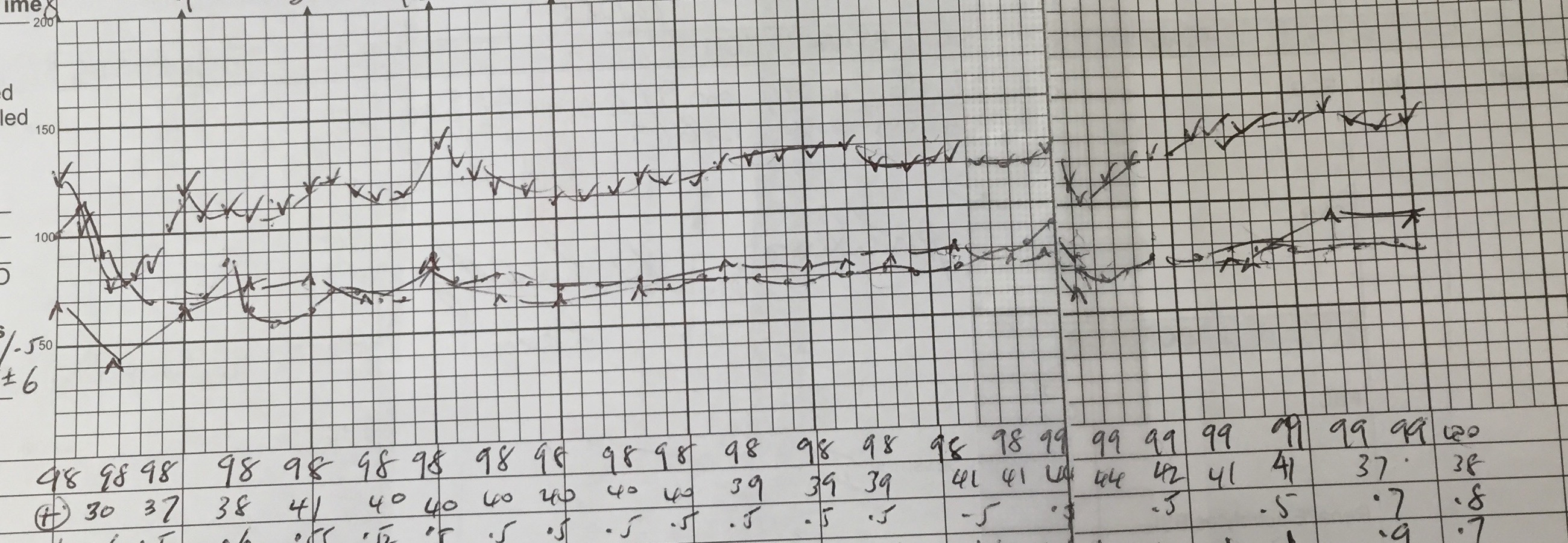
Blood pressure moves up and down, even under anesthesia.

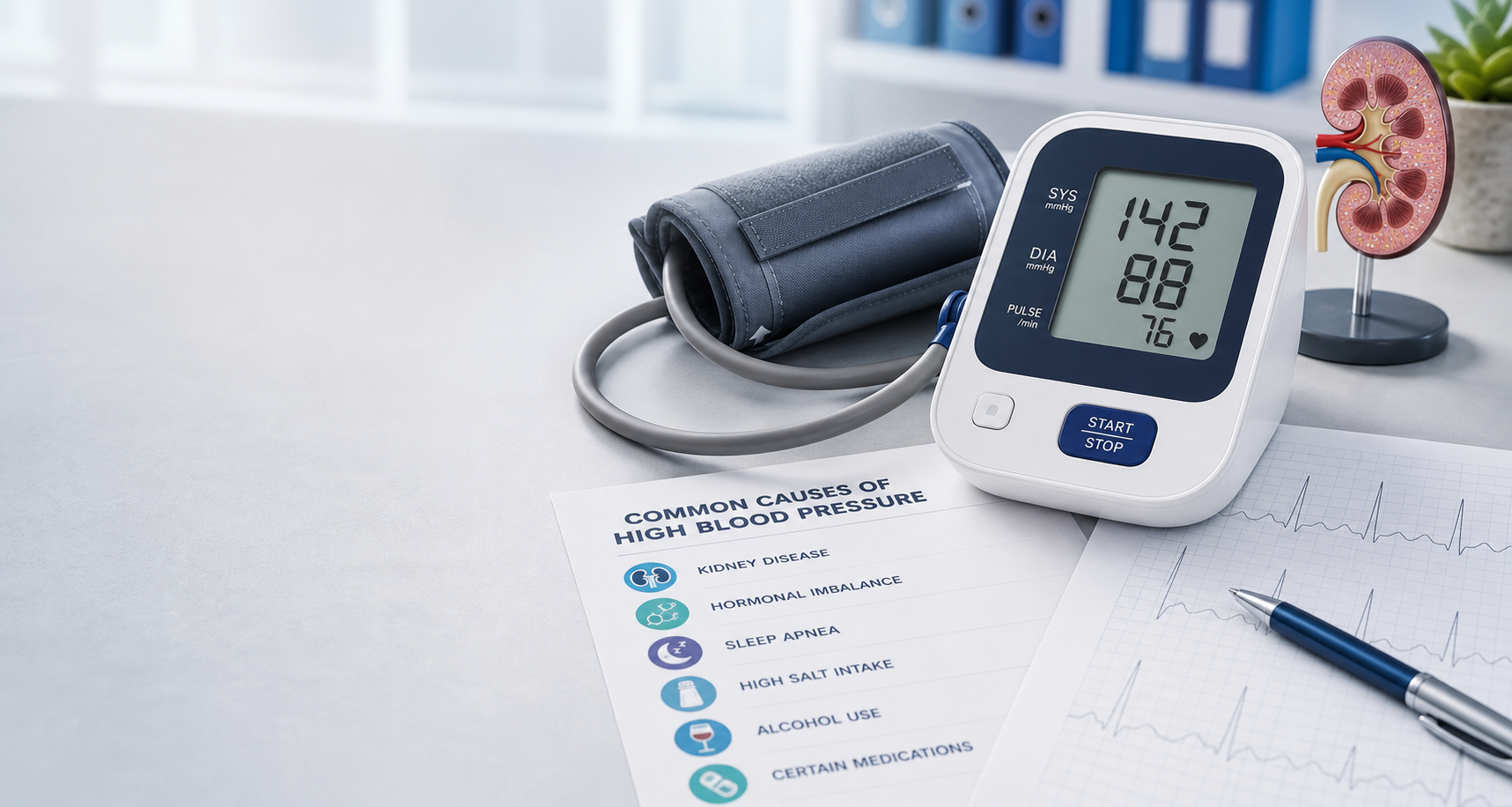
A blood pressure reading is a snapshot, not a permanent label. During a long operation, anesthetists chart blood pressure repeatedly and the trace still rises and falls with anesthesia depth, medicines, fluid shifts, bleeding, surgical stimulation, positioning, and recovery. Awake blood pressure also varies with posture, pain, exercise, stress, caffeine, alcohol, sleep, meals, a full bladder, and measurement technique.

That is why clinicians usually care about the pattern: repeated readings, home averages, ambulatory monitoring, and whether high numbers are linked with organ strain. One surprising reading deserves attention; a repeated pattern deserves a plan.

Intraoperative anesthetic charts make visible the same physiologic principle behind outpatient BP interpretation: pressure is dynamic. Induction, nociceptive stimuli, anesthetic depth, ventilation, volume status, blood loss, vasoactive agents, neuraxial effects, temperature, and surgical phase can move SBP/DBP substantially over minutes.

For chronic hypertension, avoid overinterpreting isolated values. Use standardized technique, average readings, HBPM/ABPM where useful, timing context, medication adherence, orthostatic symptoms, nocturnal pattern, and end-organ phenotype to decide whether the patient has sustained hypertension, white-coat effect, masked hypertension, or labile BP on top of true risk.

Cropped anesthetic record showing blood pressure changing repeatedly during a long surgery.
Long-surgery anesthetic record. The upper and lower BP traces move throughout the case. The lesson for everyday readings is similar: interpret blood pressure as a repeated pattern, not a single isolated number.
Body stateSleep, pain, stress, temperature, posture, hydration, and activity.
SubstancesCaffeine, nicotine, alcohol, stimulants, decongestants, and NSAIDs.
TechniqueCuff size, arm height, talking, rest time, and device validation.
PatternAverages and context are more meaningful than one number.

Diagnosis

What clinicians usually check.

Confirm hypertension, stage risk, identify organ injury, and screen for secondary causes when the phenotype asks for it.

Repeated BP readings

Clinic, home, or 24-hour ambulatory measurements help tell whether pressure is persistently high.

History and medicines

Sleep, diet, alcohol, tobacco, pain medicines, decongestants, family history, pregnancy, kidney disease, and symptoms all matter.

Blood and urine tests

Kidney function, potassium, sodium, glucose or A1c, cholesterol, urine protein, and thyroid testing may be checked.

Heart and vessel checks

An ECG, eye exam, kidney tests, and sometimes echocardiography or vascular imaging look for pressure-related damage.

Confirmation

Use average office BP across visits and out-of-office confirmation when possible. Classify by the higher SBP/DBP category and assess measurement quality.

Baseline risk

ASCVD risk, diabetes, CKD/UACR, lipids, smoking, obesity, OSA risk, family history, pregnancy potential, and prior CAD/stroke/HF/PAD guide intensity.

Core tests

BMP/CMP, creatinine/eGFR, electrolytes, fasting glucose or HbA1c, lipid profile, urinalysis/UACR, CBC when indicated, TSH selectively, ECG, and fundoscopic/echo evaluation by risk.

Secondary screen

Screen for primary aldosteronism, CKD/renovascular disease, OSA, endocrine causes, coarctation, and drug-induced hypertension when onset, severity, resistance, or labs suggest it.

Risk vs level

Risk rises with the numbers, but the person around the numbers matters too.

Blood pressure risk is continuous: higher usual pressure generally means higher risk. A stage 1 reading in a smoker with diabetes and kidney disease is not the same as the same reading in an otherwise low-risk adult.

Hypertension is a major public and personal health issue because it is easy to ignore until damage is already visible: kidney damage, stroke, hypertensive heart disease with left ventricular hypertrophy, heart failure, coronary disease, and myocardial infarction. The quiet years are often the best years to intervene.

Elevated BP

Often the best window for prevention. Salt reduction, weight change, activity, sleep, and alcohol reduction can keep it from becoming sustained hypertension.

Stage 1

Management depends on overall cardiovascular risk, prior disease, kidney disease, diabetes, and whether repeated readings confirm the pattern.

Stage 2

Risk and organ strain are higher. Many adults need medication plus lifestyle treatment, with timely follow-up and dose adjustment.

Severe range

Symptoms or acute organ injury can mean hypertensive emergency. Without those, urgent clinician contact is still needed.

HeartHeart attack, heart failure, left ventricular hypertrophy, atrial fibrillation.
BrainIschemic stroke, brain bleed, vascular cognitive impairment.
KidneysChronic kidney disease, albuminuria, kidney failure progression.
VesselsAortic disease, peripheral artery disease, eye vessel damage.

Eye exam clues

Retinal changes your optometrist may notice.

The retina is one of the few places where small blood vessels can be seen directly. Long-standing or severe high blood pressure can leave clues such as narrowed retinal arteries, arteriovenous nicking, flame-shaped hemorrhages, cotton-wool spots, hard exudates, swelling, or optic disc swelling.

These findings do not prove the exact cause by themselves. Diabetes, vein occlusion, cholesterol disease, anemia, inflammation, and other eye conditions can overlap. But when an optometrist sees vascular retinal changes, it is a good reason to check blood pressure, diabetes risk, kidney disease, cholesterol, and overall cardiovascular risk.

Hypertensive retinopathy ranges from chronic arteriolar narrowing, AV crossing changes, and copper/silver wiring to acute retinal hemorrhages, cotton-wool spots, hard exudates, macular edema, and optic disc edema in severe hypertension. Moderate to severe retinopathy should prompt systemic BP assessment, target-organ review, diabetes/CKD evaluation, medication review, and urgency triage.

Retinal findings are not disease-specific. Correlate with duration and severity of hypertension, diabetes status, renal disease, neurologic symptoms, pregnancy/postpartum state, and ocular differential diagnoses such as diabetic retinopathy, retinal vein occlusion, anemia/leukemia, inflammatory vasculitis, and papilledema from other causes.

Retinal photograph with surrounding examples of systemic diseases and cardiovascular risks associated with retinal imaging.
Retinal imaging as a vascular window. The eye exam can reveal pressure-related microvascular damage and can also raise broader questions about diabetes, kidney disease, cholesterol, stroke, and heart risk.
Chronic pressureArteriolar narrowing, AV nicking, copper or silver wiring.
More active injuryFlame hemorrhages, cotton-wool spots, hard exudates, retinal edema.
Severe warningOptic disc swelling or vision symptoms can signal urgent disease.
Next stepConfirm BP and assess diabetes, kidneys, cholesterol, brain, and heart risk.

Briefly

Symptoms and treatments, kept short for this causes site.

Most high blood pressure has no symptoms. Possible symptoms in severe hypertension can include headache, nosebleed, vision changes, chest pain, breathlessness, confusion, weakness, or trouble speaking, but symptoms are not a reliable way to detect it.

Symptoms should trigger assessment for hypertensive emergency, ACS, acute HF/pulmonary edema, stroke/TIA/ICH, aortic syndrome, AKI, preeclampsia/eclampsia, and drug/toxin states. Treatment depends on urgency, organ injury, comorbidity, pregnancy status, and chronic regimen.

Lifestyle foundations

DASH-style eating, lower sodium, more potassium-rich foods when safe, regular activity, weight reduction when appropriate, sleep apnea care, less alcohol, and no tobacco.

Medicines

Common first-line classes include thiazide-type diuretics, ACE inhibitors or ARBs, and calcium channel blockers. Choice depends on age, kidney disease, diabetes, heart disease, pregnancy potential, side effects, and other medicines.

Follow-up

Successful treatment usually means repeat measurements, dose adjustments, side-effect checks, lab monitoring, and looking again for secondary causes when control is difficult.

BP images

Standard blood pressure visuals.

Digital blood pressure monitor showing 142 over 88 beside a cuff and kidney model.
Home-style digital monitor. Upper-arm devices are commonly used for home blood pressure tracking when validated and correctly fitted.
Clinician checking blood pressure with a cuff and stethoscope.
Manual measurement. A cuff and stethoscope can measure brachial blood pressure when technique is careful. Image from Wikimedia Commons.
Aneroid sphygmomanometer and stethoscope.
Sphygmomanometer. Mechanical gauges remain a standard symbol of blood pressure measurement. Source: CDC via Wikimedia Commons.
Blood pressure cuff being used on an arm.
Cuff placement. Correct cuff size and arm position are central to reliable readings. Image by Sistema d'Emergencies Mediques, CC BY-SA IGO 3.0.
Retinal photograph surrounded by labels for systemic disease research and cardiovascular prediction.
Retinal vascular clues. Optometrists and eye doctors may see signs that support checking blood pressure and broader vascular risk.
Anesthetic record showing blood pressure varying during surgery.
BP variation over time. Surgical anesthetic records show what home logs also reveal: blood pressure moves, so repeated patterns matter.

Languages

Country language presets and full-page translation.

When this site is live, use the selector to open the page through Google Translate. The country presets are quick links for languages commonly needed in cardiovascular care.

Common languages